STEMCELL Technologies MyoCult MyoCult SF Expansion Supplement Kit (Human)
MyoCult Expansion kit (Human) (ST-05960)の後継品です
- 研究用
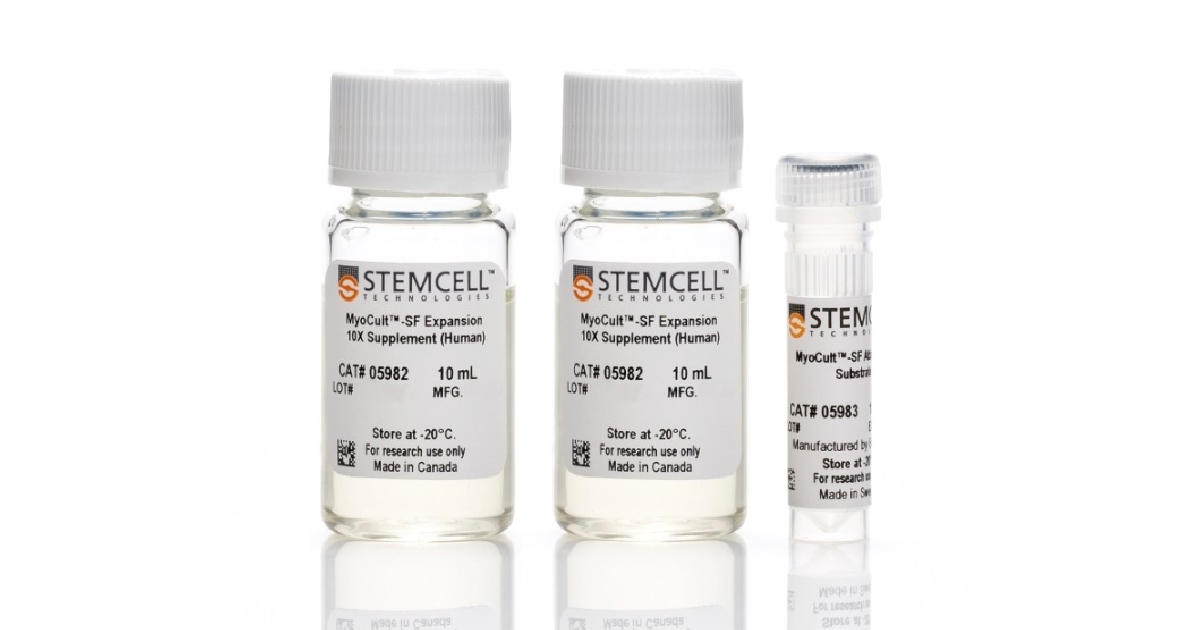
MyoCult™-SF Expansion Supplement Kit (Human) (ST-05980)は、初代ヒト骨格筋前駆細胞(筋芽細胞)およびヒト多能性幹細胞(hPSC)由来の筋芽細胞を無血清条件で培養するキットです。
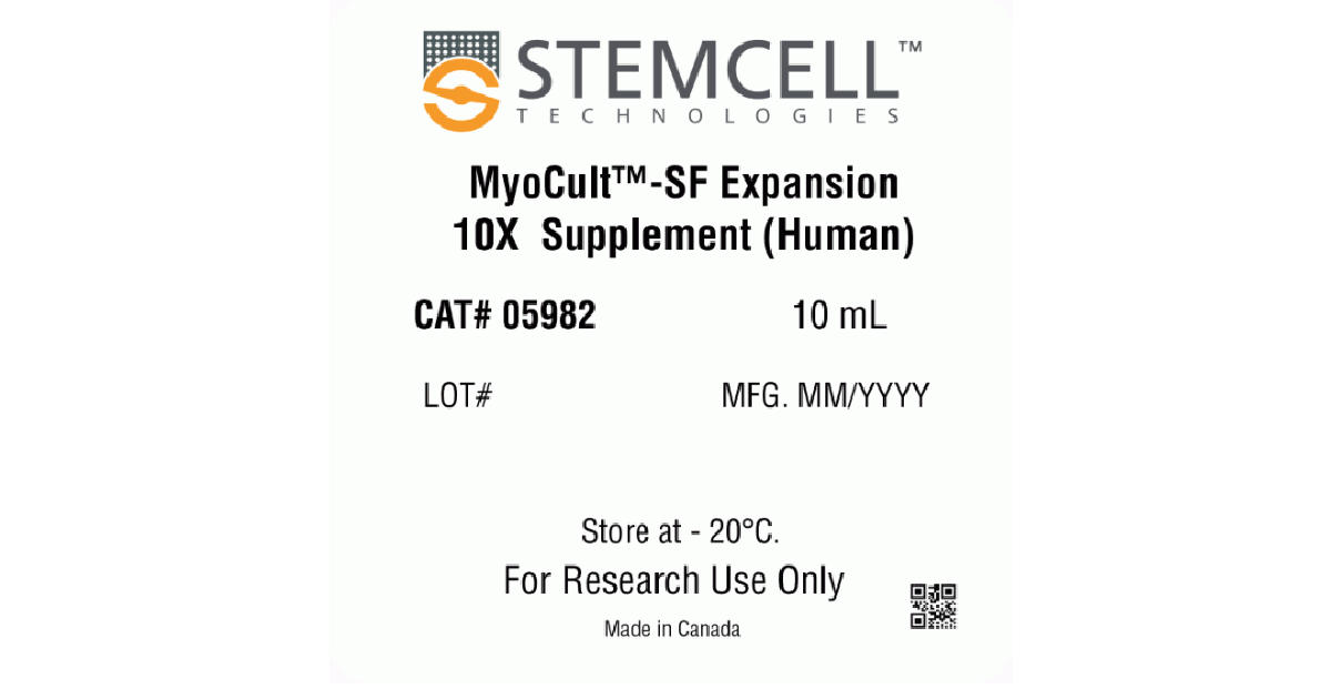
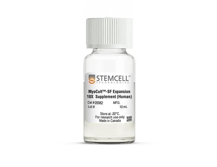
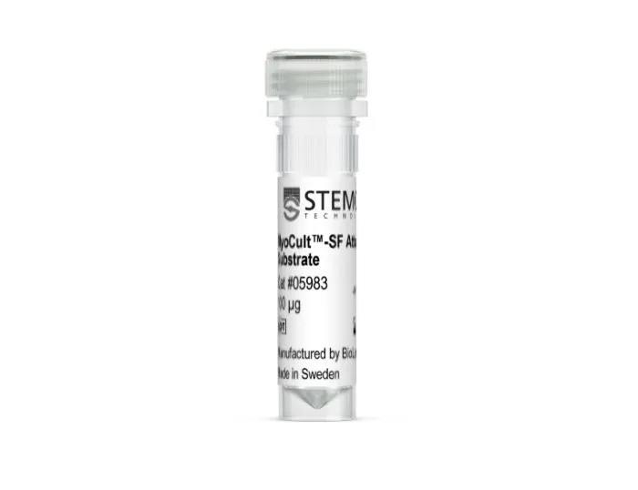
本品には、筋芽細胞のin vitro での誘導と増殖に最適化されたサプリメント MyoCult™-SF Expansion 10X Supplement(ST-05982)、および細胞接着基質 MyoCult™-SF Attachment Substrate(ST-05983)が含まれています。サプリメントは、基礎培地(DMEM with 1000 mg/L D-glucose [例:ST-36253]、別売)に添加して、MyoCult™-SF Expansion Mediumを調製する必要があります。細胞接着基質は、培養する細胞に応じてオプションで使用します。各コンポーネントは、個別に購入することもできます。
初代筋芽細胞を培養する場合、培養器具をMyoCult™-SF Attachment Substrateでコーティングすることが推奨されます。hPSC由来の筋芽細胞を培養する場合、Corning® Matrigel® hESC-Qualified Matrixでコーティングすることができます。
MyoCult™-SF Expansion Mediumで培養した筋芽細胞は、MyoCult™ Differentiation Kit (Human)(ST-05965)で培養して多核筋管へと分化できます。
製品の特長
MyoCult™-SF Expansion Supplement Kit (Human)で、ヒト筋芽細胞を誘導・増殖
- ヒト骨格筋前駆細胞の無血清条件での誘導および長期増殖をサポート
- 骨格筋前駆細胞の分化能を保ったまま増殖
- 徹底した原材料スクリーニングと品質管理により、ロット間差を最小限に抑制
- 拡大培養した骨格筋前駆細胞は、MyoCult™ Differentiation Kitで分化可能
筋芽細胞のアプリケーション
- 筋形成の生物学的、電気生理学的な研究
- 遺伝性筋疾患(筋ジストロフィー)モデル
- 筋関連のがん(横紋筋肉腫)モデル
- 骨格筋疾患(悪液質、糖尿病、代謝性疾患)モデル
- 創薬スクリーニングのプラットフォーム
- 共培養システム
ヒト筋芽細胞の増殖・分化の流れ
Human skeletal muscle tissue is digested into a single cell suspension and plated into MyoCult™-SF Expansion Medium. Myoblasts are then enriched by cell sorting or using a selection kit such as the EasySep™ Human CD56 Positive Selection Kit (Catalog #17855). Purified myoblasts are then culture-expanded or differentiated into myotubes for further studies. Myoblasts can be derived and expanded, using the MyoCult™-SF Expansion Kit (Catalog #05980) and then differentiated using the MyoCult™ Differentiation Kit (Catalog #05965).
データ紹介
ヒト骨格筋組織からの骨格筋前駆細胞の誘導
Myogenic progenitor cells were derived from human skeletal muscle tissue and culture-expanded using the MyoCult™-SF Expansion Kit or a serum-containing medium. (A) Following 6 days of expansion, myoblasts were fixed and immunostained for PAX7 (green), a thymine analogue (EdU; red) and nuclei (DAPI; blue). Total percentage of cells expressing (B) PAX7 or (C) PAX7 and EdU were quantified (n = 4). Data was generated and used with permission by Dr. Penny M. Gilbert’s Lab, Department of Biochemistry, University of Toronto.
筋芽細胞の長期増殖

Myoblasts were culture-expanded in MyoCult™-SF Expansion Medium or in serum-containing medium.
(A) Myoblasts culture-expanded in MyoCult™-SF Expansion Medium displayed superior expansion rate than when cells were expanded in serum-containing medium (n=3).
(B) Expansion of myoblasts in MyoCult™-SF Expansion Medium generates a greater yield of CD56+ cells after 9 passages compared to myoblasts cultured in serum-containing medium (P9; n = 2).
Data was generated and used with permission by Dr. Penny M. Gilbert’s Lab, Department of Biochemistry, University of Toronto.
増殖後の骨格筋前駆細胞の分化能保持と移植

Myoblasts derived and expanded in MyoCult™-SF Expansion Medium were further differentiated into myotubes using the MyoCult™ Differentiation Kit (Catalog #05965) at passage 5.
(A) Myotubes differentiated from myoblasts were immunostained for myosin heavy chain (MyHC; red) and nuclei (DAPI; blue).
(B) Fusion index displaying approximately 60% of total nuclei were found within myotubes expressing MyHC. Each donor is indicated by a yellow square. Data from 5 independent donors.
iPS細胞由来筋芽細胞の増殖

(A) iPSC-derived myogenic progenitor cells displayed long-term expansion when using the MyoCult™ Expansion Kit. iPSC-derived myogenic progenitor cells were generated using Protocol A (Chal et al. from Nature Protocols (2016)) and Protocol B (Xi et al. Cell Rep (2017)).
(B) Culture-expanded iPSC-derived myogenic progenitor cells expressed MyHC (red) and MyoD (green).


