STEMCELL Technologies STEMdiff STEMdiff Pancreatic Progenitor Kit
- 研究用
STEMdiff™ Pancreatic Progenitor Kit(ST-05120)は、ヒト多能性幹細胞(hPSC)から膵前駆細胞を効率的に再現性高く作製する無血清培地です。
細胞は4つの段階すなわち、1)胚体内胚葉、2)原始腸管、3)後部前腸内胚葉、4)膵前駆細胞、を経て分化します。分化した細胞は、PDX-1、NKX6.1およびSOX9を含む膵前駆細胞の重要なマーカーを発現し、インスリンとグルカゴンの発現が上昇します。得られた膵前駆細胞はさらに、インスリン産生ベータ細胞または他の内分泌および外分泌膵臓細胞に分化させることができます。
本品は、mTeSR™1(ST-85850)またはmTeSR™ Plus(ST-100-0276)で維持されたhPSCからの分化に最適化されています。

2018/05/14 12:00の製品情報

本製品は研究目的にのみ使用し、人や動物の医療用・臨床診断用・食品用としては使用しないようにご注意ください。
製品の特長
STEMdiff™ Pancreatic Progenitor Kitで、hPSCから膵前駆細胞へと無血清で分化誘導
- 動物モデルへの移植、またはインスリン産生β細胞への成熟が可能な、機能性の膵前駆細胞を作製
- 複数のヒトES/iPS細胞株から、高い効率と再現性でPDX-1、NKX6.1、およびSOX9を発現する膵前駆細胞に分化
- 無血清培地
- mTeSR™1またはmTeSR™ Plusで維持された複数のヒトES/iPS細胞株で、膵前駆細胞分化を確認済み
使用方法
hPSC由来膵前駆細胞の作製プロトコル概要
ステージ1-4を経て14日間で分化します。詳細な使用方法は、製品添付文書をご覧ください。
データ紹介
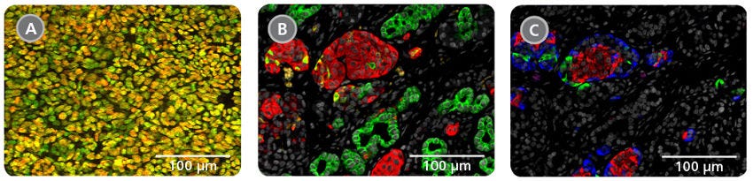
Figure 1. Pancreatic Progenitor Cells can Mature into Endocrine and Exocrine Cells
A) Representative image showing pancreatic progenitor cells expressing PDX-1 (red) and NKX6.1 (green). Yellow staining indicates co‑expression of both markers in the majority of cells as is observed in the developing human pancreas. (B, C) Cells transplanted into mice can mature into endocrine and exocrine cells.
B) shows endocrine clusters expressing synaptophysin (red) surrounded by ductal structures expressing CK-19 (green).
C) Shows islet-like structures containing monohormonal cells that individually express insulin (red), glucagon (green) or somatostatin (blue). Data in (B, C) are from the laboratory of Dr. Timothy J. Kieffer (University of British Columbia, Vancouver, Canada).
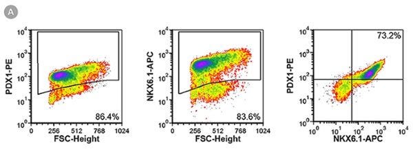
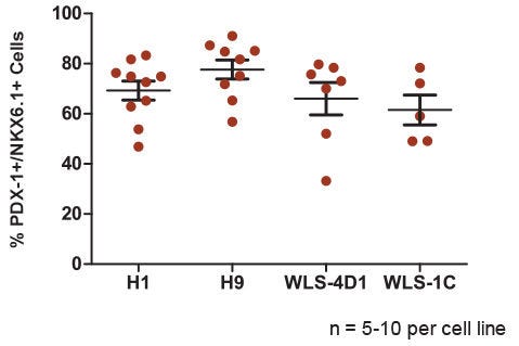
Figure 2. The STEMdiff™ Pancreatic Progenitor Kit Functions Efficiently Across Multiple hPSC Lines
PDX-1 and NKX6.1 expression measured in pancreatic progenitor cells derived from four different hPSC lines (H1, H9, WLS-4D1 and WLS-1C) at the end of Stage 4. (A) Representative flow cytometry plots show PDX-1 and NKX6.1 co-expression in differentiated H9 cells. (B) Quantitative data for PDX-1/NKX6.1 co-expression in two human ES (H1 and H9) and two human iPS (WLS-4D1 and WLS-1C) cell lines (n = 5-10 per cell line). Data are plotted as individual points representing the mean of duplicates within a single experiment. The horizontal line represents the mean of all experiments, with error bars indicating the standard error of the mean (SEM). The average efficiency of pancreatic progenitor differentiation ranges from 61.5% to 77.7% depending on the cell line.
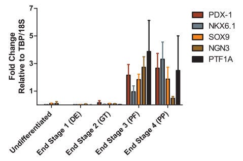
Figure 3. Gene Expression Profile is Indicative of Transition to Pancreatic Progenitor Cells
Gene expression profile at the end of each stage of differentiation for key markers of pancreatic progenitor cells. Expression was normalized to 18S ribosomal RNA and TATA Binding Protein (TBP). Data are the mean ± SEM for 3 - 5 experiments. Expression pattern is consistent with published data.²
1. Riedel M et al. (2012) Immunohistochemical characterization of cells co-producing insulin and glucagon in the developing human pancreas. Diabetologia 55(2): 372-81.
2. Rezania A et al. (2014) Reversal of diabetes with insulin-producing cells derived in vitro from human pluripotent stem cells. Nat Biotechnol 32(11): 1121-33.
Figure 4. Generation of Pancreatic Progenitors from hPSCs Maintained in mTeSR™ Plus
(A) Representative density plots showing PDX-1 and NKX6.1 expression in cells cultured in mTeSR™1 (daily feeds) or mTeSR™ Plus (restricted feeds), following differentiation using the STEMdiff™ Pancreatic Progenitor Kit. (B) Quantitative analysis of pancreatic progenitor formation in multiple hPS (H9, STiPS-M001, WLS-1C) cell lines maintained with mTeSR™1 or mTeSR™ Plus as measured by co-expression of PDX-1 and NKX6.1. Data are expressed as the mean percentage of cells (± SEM) expressing both markers; n=3.


