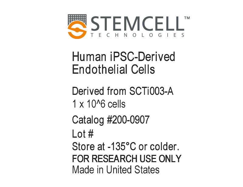
STEMCELL Technologies STEMdiff Human iPSC-Derived Endothelial Cells
- 研究用
- 新製品
Human iPSC-Derived Endothelial Cells は、ただちに使用可能な高品質のヒト内皮細胞(endothelial cells:ECs)です。本品は、健康な女性ドナー由来のヒト人工多能性幹細胞(iPSC)コントロールラインであるSCTi003-AからSTEMdiffTM Mesoderm Induction MediumとSTEMdiffTM Endothelial Differentiation Kitを使用して分化させ、凍結保存された内皮細胞です。分化した細胞からスタートすることで、研究に必要な大量の細胞を迅速に得られ、自信をもってワークフローを開始することができます。
STEMdiffTM Endothelial Expansion Culture Kitを用いて解凍したECは、最低3回の継代培養により増殖能を維持しながら実験規模を拡大することができます。拡大培養した細胞は、内皮特異的マーカーであるCD31、CD144、CD309 (VEGFR2)を高発現し、造血系(CD45)および上皮系(CD326)マーカーは不検出の状態を維持します。
本品は研究用製品(RUO)で、学術・商業研究用としての承認を受けています。血液サンプルは、治験審査委員会(IRB)またはその他の規制当局が承認した同意書およびプロトコルを用いて倫理的に提供されています。
2018/05/14 12:00の製品情報

本製品は研究目的にのみ使用し、人や動物の医療用・臨床診断用・食品用としては使用しないようにご注意ください。
製品の特長
ヒトiPS細胞株 SCTi003-Aから分化した内皮細胞(EC)
- 解凍直後から高品質なヒト内皮細胞を取得可能
- STEMdiff™ Endothelial Expansion Culture Kitで増殖能と品質を維持しながら容易に細胞を拡大培養可能
- 詳細に特性解析済みのヒトiPSCコントロール株SCTi003-A由来の高純度ECで研究開始、実験の再現性を確保
ご注意
ソースセルバンクのドナー詳細および細胞品質特性については、以下のSCTi003-Aの情報をご参照ください。
コントロールに最適な凍結ヒトiPS細胞株
SCTi003-Aは、αβT細胞に由来し、VDJ配列の再構成を受けています。核型的に安定であり、3胚葉分化能を示し、未分化細胞マーカーを発現し、非組み込み型技術によりリプログラミングされています。hPSCreg®に登録されており、コミュニティ基準に基づく倫理的・生物学的適合性が保証されます。詳細については、ロット別の検査証明書(CoA)およびiPSC株に関するよくある質問もご参照ください。
データ紹介
Figure 1. Human iPSC-Derived Endothelial Cells Exhibit High-Quality Morphology and Robust Expansion for Multiple Passages
(A) Representative microscopy image showing cryopreserved human iPSC-derived endothelial cells thawed and maintained in STEMdiff™ Endothelial Expansion Medium for 4 days and ready for passaging at ~90 - 100% confluency.
(B) Cells show robust expansion post-thaw. Data points show the fold increase over 4 passages post-thaw from one technical replicate.
(C) Human iPSC-derived endothelial cells were thawed and maintained in STEMdiff™ Endothelial Expansion Medium. Cells were passaged every 3 - 5 days post-thaw, and cell expansion was analyzed as cumulative cell number over 4 passages from one technical replicate.
Figure 2.Human iPSC-Derived Endothelial Cells Express Characteristic Markers
Human iPSC-derived endothelial cells were thawed, cultured in STEMdiff™ Endothelial Expansion Medium for four passages, and characterized using flow cytometry for endothelial markers. The cells show high expression of (A) CD31/CD144, (B) CD309, and (C) CD105. (D) A representative flow cytometry plot shows that the cells were negative for the epithelial marker CD326 (EpCAM).
Figure 3. Human iPSC-Derived Endothelial Cells Are Functional and Mature
Human iPSC-derived endothelial cells were thawed and cultured in STEMdiff™ Endothelial Expansion Medium for two passages before assessing functionality and maturity.
(A) Immunohistochemistry confirmed the expression of endothelial markers CD31 (green), VWF (magenta), and DAPI (gray), and (B) the ability to take up acetylated LDL (green) and Hoechst 33342 (Blue).
(C) Cells were able to form tubular networks in vitro in a tube formation assay when plated on Corning® Matrigel® for 24 hours.

