STEMCELL Technologies PancreaCult PancreaCult Organoid Medium (Mouse)
- 研究用
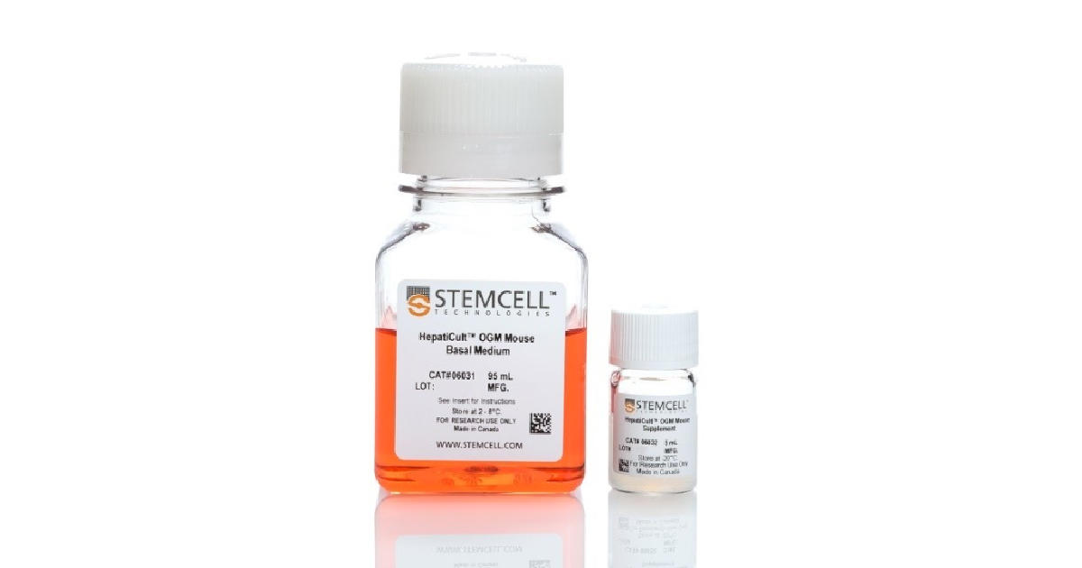
PancreaCult™ Organoid Growth Medium (Mouse)は、マウス膵外分泌オルガノイドを樹立し維持するための、組成が明確 (defined) な無血清培地です。
これらの膵臓オルガノイドあるいは「ミニ膵臓」は、膵臓の細胞生物学、疾患、がんの研究向けの in vitro 器官培養系となります。PancreaCult™で培養したオルガノイドは、膵臓幹細胞 (LGR5)、前駆細胞 (PDX1、SOX9)、および膵管細胞 (CAR2、MUC1、KRT19) の各マーカー遺伝子を発現する上皮を特徴とします。膵オルガノイドは、3〜6日ごとに継代して長期に維持でき、凍結保存も可能です。
PancreaCult™では、マウス膵オルガノイドをCorning® Matrigel®のドームに埋め込んで、または希釈Matrigel®に懸濁して培養できます。オルガノイド培養は、生理的に適切で便利な in vitro 系で膵臓上皮を特徴付けることを可能にし、動物使用の必要性を減らします。
本品はHUBとの共同開発製品です。商業目的で使用する場合は、HUB(www.huborganoids.nl/)へご連絡の上、商用ライセンスまたはHUBとのライセンスに関する説明を受けてください。
製品の特長
PancreaCult™ Organoid Growth Medium (Mouse) で、マウス膵外分泌オルガノイドを樹立・維持
特長
- 簡易な in vitro 培養系:7日以内で膵臓オルガノイドを樹立
- STEP-BY-STEPの詳細なプロトコル:膵障害モデル、膵管の手動単離、幹細胞ソーティングは不要
- シンプルな2-コンポーネント組成:成分が明確な無血清培地
- フレキシブルなプロトコル:マトリックスドームまたは懸濁液でのオルガノイド培養に対応
用途
- 膵幹細胞・膵管上皮細胞研究
- 膵臓がん・疾患モデル研究
- 膵臓移植研究
- 創薬毒性試験
データ紹介
成長中のマウス膵外分泌オルガノイド

Pancreatic exocrine organoids are observed within one week when cultured in (A) Corning® Matrigel® domes or (B) a dilute Matrigel® suspension. Organoids were imaged during passage 2, on day 4.
さまざまな試料から培養開始できるマウス膵外分泌オルガノイド

PancreaCult™ Organoid Growth Medium (Mouse) enables the initiation of pancreatic exocrine organoids from (A) duct fragments, (B) single cells and (C) cryopreserved organoid fragments. All organoids were grown in Matrigel® domes. Organoids were imaged on day 4 or day 5 of primary culture (duct fragments and single cells, respectively) or day 3 of the first passage post-thaw (cryopreserved organoids).
マトリゲルのドームまたは懸濁液で培養可能
Organoids cultured using PancreaCult™ Organoid Growth Medium (Mouse) from freshly isolated pancreatic tissue fragments and plated in (A) Matrigel® domes or (B) as a dilute Matrigel® suspension. Organoids grown in either culture condition are typically ready for passage within 3 - 6 days.
膵臓前駆細胞と膵管細胞のマーカーを発現

Pancreatic exocrine organoids grown in PancreaCult™ and stained for nuclei (DAPI, blue), ductal marker KRT19 (green) and pancreatic progenitor marker PDX1 (red). Organoids were imaged during passage 12 on day 5.
Note: The folded appearance of epithelium is a function of cryosectioning and not representative of the shape of proliferating organoids.
継代中に膵臓マーカー発現を保持

Pancreatic organoids express stem cell markers and those typical of the pancreatic exocrine system, including (A) Axin2, (B) Krt19, (C) Muc1 and (D) Pdx1. Relative quantification (RQ) of each marker is reported relative to the 18S and TBP housekeeping genes and normalized to C57/Bl6 pancreatic tissue. Marker expression was assayed during early passages (passage 1-5) and late passages (passage 6-10).
継代によるオルガノイドの増殖

Organoids cultured with PancreaCult™ Organoid Growth Medium (Mouse) show efficient growth over multiple passages. Cultures were split with an average split ratio of 1:16 at each passage.
膵臓がん由来の膵外分泌オルガノイド

PancreaCult™ Organoid Growth Medium (Mouse) supports the growth of organoids from pancreatic carcinomas. Pancreatic ducts were isolated from KPC mice (Kras+/LSL-G12D; Trp53+/LSL-R172H; Pdx1-Cre) and cultured in PancreaCult™ Organoid Growth Medium (Mouse). Organoids were imaged on (A) day 4 of primary culture and (B) day three after the first passage. An activated KRAS genotype was retained in organoids during culture. Data used with permission from Dr. David Tuveson.


