STEMCELL Technologies STEMdiff STEMdiff-ACF RPE Differentiation Kit
- 研究用
- 新製品
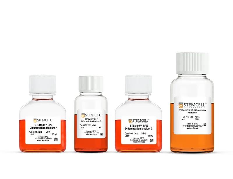
STEMdiff-ACF™ RPE Differentiation Kit(ST-100-1367)は、ヒト多能性幹細胞(hPSC)から未熟な網膜色素上皮(retinal pigment epithelium; RPE)を14日間で迅速かつ堅牢に作製できます。本品は動物性成分不含および無血清であり、mTeSR™ 1またはmTeSR™ Plus培地中でクランプ(凝集)培養して維持されたhPSCから未熟なRPE(>50% PMEL17+)を作製します。
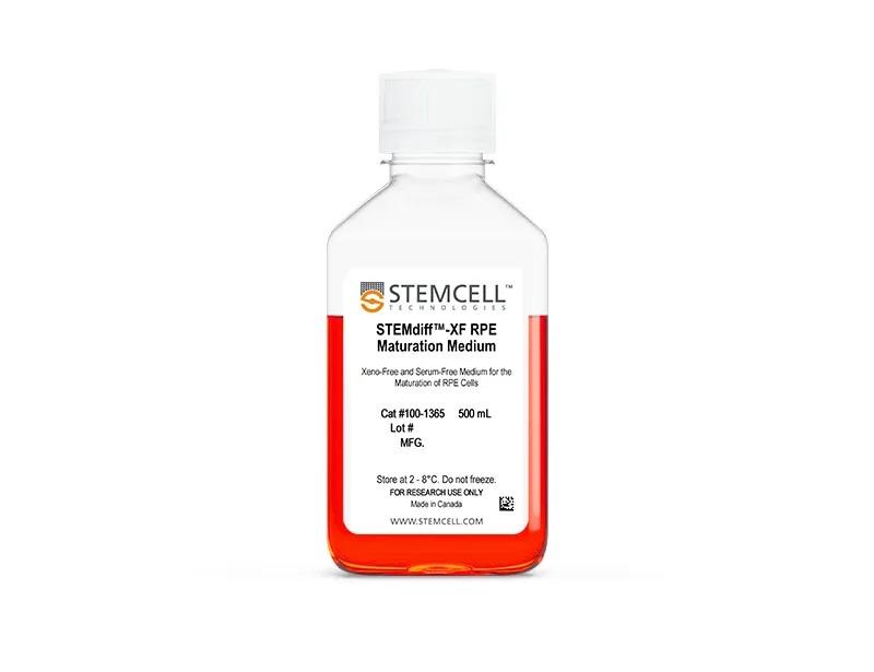
本品で作製した未成熟RPEは、中間的な細胞バンクとして凍結保存することも、STEMdiff-XF™ RPE Maturation Medium(ST-100-1365)でさらに成熟させ機能的なRPEにすることもできます。両製品の併用によって、hPSCから高純度のRPE(以下のデータ紹介参照)をわずか49日で、手作業による選択や細胞濃縮を行わずに作製できます。RPE細胞分化・成熟における収量と生存率を向上させるには、このワークフローにSTEMdiff-ACF™ RPE Plating Supplement(ST-100-1364)を追加することができます。
これらの製品を使用して得られたRPEは、ヒト網膜の発達と疾患のモデル化、薬物スクリーニング、細胞・遺伝子治療の検証、および高次元な組織モデル開発に使用できます。
本品を商用または臨床用途で使用する場合は、弊社までお問い合わせください。
【関連製品】本品を用いて作製された、ヒトiPS細胞由来のRPE細胞はこちら>>
製品の特長
STEMdiff™-ACF RPE Differentiation Kitで、ヒト多能性幹細胞から未成熟な網膜色素上皮を作製
- 動物性成分も血清も含まない培地組成で、一貫性のある結果を取得
- シンプルで培養規模を変更可能なワークフローで、効率的にRPE分化
- hPSCから未成熟RPE細胞をわずか14日で作製
- 複数のヒトESおよびiPS細胞株で、高純度かつ機能的なRPEへの分化を検証済み
- 便利でユーザーフレンドリーな信頼できるワークフロー
データ紹介
Robust and Rapid Generation of Mature Retinal Pigment Epithelial Cells (RPE) across multiple PSC Cell Lines with the STEMdiff™-ACF RPE Differentiation Kit
hPSCs were cultured for 14 Days using STEMdiff™-ACF RPE Differentiation Kit and subsequently subcultured in STEMdiff™-XF RPE Maturation Medium. Flow cytometry expression of RPE markers are shown at Day 14 and Day 49. (A) The percentage of cells expressing PMEL17, RPE65, EZRIN, and CRALBP and (B) Viable cell yields for 4 hPSC cell lines. PMEL17 is expressed at Day 14 and 49 while the other markers are only present at Day 49. Data are reported as mean + SEM; n = 16 -20. (C) A cell pellet of mature RPE cells demonstrates the pigmentation. Maturation is further demonstrated with immunohistochemistry for expression of RPE markers at Day 49. (D, E, F, G) Mature RPE display tight junctions marked by localization of ZO1 and BEST1 to cell junctions. Mature RPE are polarized, expressing EZRIN apically and ZO1 subapically and express proteins required for the visual cycle (RPE65).
Mature Retinal Pigment Epithelial (RPE) Cells Display Key Functionalities Corresponding to RPE Behaviour
hPSC’s were cultured for 14 Days using STEMdiff™-ACF RPE Differentiation Kit and subsequently subcultured on cell culture inserts in STEMdiff™-XF RPE Maturation Medium for 5 weeks. Apical and basal conditioned medium were collected from Mature RPE, and a sandwich ELISA was performed to quantify Vascular Endothelial Growth Factor (VEGF) and Pigment Epithelial Derived Growth Factor (PEDF) secretion. (A, B) Mature RPE secreted more basal VEGF and apical PEDF demonstrating RPE display correct apicobasal polarity. Data shown as mean + SEM; n = 3. (C) Mature RPE were able to generate a strong barrier with high transepithelial resistance (TER). Data shown as mean + SEM; n = 3-6. (D) Mature RPE were fed FITC-labelled bovine photoreceptor outer segments (POS) for 4 to 5 hours prior to being enzymatically dissociated for flow cytometry analysis or fixed with paraformaldehyde for immunostaining. (E) Mature RPE efficiently internalize bovine POS. Data shown as mean + SEM; n = 3. (F) A cross-sectional schematic of the cell insert culture system.


